


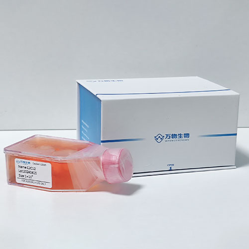
人淋巴管內(nèi)皮細(xì)胞 Human Lymphatic Endothelial Cells
目錄價(jià)
¥ 0.00
 一鍵復(fù)制產(chǎn)品信息
別稱HLEC;Human Lymphatic Endothelial Cells
貨號(hào)Delf-16791
規(guī)格


產(chǎn)品推薦


聯(lián)系方式
- 電話:400-1016-218
- 郵箱:355185756@qq.com
- 地址:安徽省合肥市高新區(qū)黃山路602號(hào)合肥國家大學(xué)科技園A401室
產(chǎn)品詳情
Cell Specification
The lymphatic system is an essential part of the immune system. It helps maintain tissue homeostasis, including interstitial protein transport, tissue fluid balance, and development of cellular immunity. Lymph nodes, which are located throughout the lymphatic system, contain large numbers of lymphocytes, macrophages and antigen presenting cells that together initiate primary immune response [1]. Specialized lymphatic endothelial cells (LEC) in the cortex assist in the primary immune response by recruiting intravascular lymphocytes as they circulate past [2]. In addition, LEC are responsible for facilitating the transmigration of intravascular lymphocytes into the reticular meshwork where the lymphocytes can interact with antigen presenting cells [2]. LEC have been shown to be associated with chronic inflammation and cancers [3].
HLEC from ScienCell Research Laboratories are isolated from human lymph node. HLEC are cryopreserved at passage one and delivered frozen. Each vial contains >5 x 10^5 cells in 1 ml volume. HLEC are characterized by immunofluorescence with antibodies specific to vWF/Factor VIII and/or CD31 (PECAM1). HLEC are negative for HIV- 1, HBV, HCV, mycoplasma, bacteria, yeast, and fungi. HLEC are guaranteed to further expand for 10 population doublings under the conditions provided by ScienCell Research Laboratories.
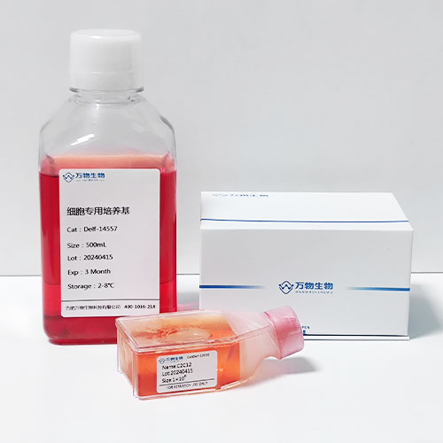
Recommended Medium
It is recommended to use Endothelial Cell Medium (ECM, Cat. #1001) for culturing HLEC in vitro.
Product Use
HLEC are for research use only. They are not approved for human or animal use, or for application in in vitro diagnostic procedures.
Storage
Upon receiving, directly and immediately transfer the cells from dry ice to liquid nitrogen and keep the cells in liquid nitrogen until they are needed for experiments.
Shipping
Dry ice.
References
[1] Kaldjian EP, Gretz JE, Anderson AO, Shi Y, Shaw S . (2001) “Spatial and molecular organization of lymph node T cell cortex: a labyrinthine cavity bounded by an epithelium-like monolayer of fibroblastic reticular cells anchored to basement membrane-like extracellular matrix. ” Int Immunol. 13: 1243-53.
[2] Willard-Mack CL. (2006) “Normal structure, function, and histology of lymph nodes.” Toxicol Pathol. 34: 409-24.
[3] Martinet L, Garrido I, Filleron T, Le Guellec S, Bellard E, Fournie JJ, Rochaix P, Girard JP. (2011) “Human solid tumors contain high endothelial venules: association with T- and B-lymphocyte infiltration and favorable prognosis in breast cancer.” Cancer Res. 71: 5678-87.
Instructions for culturing primary cells
Caution:Cryopreserved primary cells are very delicate. Thaw the vial in a 37oC water bath and return the cells to culture as quickly as possible with minimal handling! Do not centrifuge the cells after thawing as this can damage the cells.
Initiating the culture:
Note: ScienCell primary cells must be cultured in a 37oC, 5% CO2 incubator. Cells are only warranted if ScienCell media and reagents are used and the recommended protocols are followed.
1. Prepare a fibronectin-coated culture vessel (2 μg/cm2, T-75 flask is recommended). To obtain a 2 μg/cm2 fibronectin-coated culture vessel, add 10 ml of sterile Dulbecco’s phosphate buffered saline, Ca++- and Mg++-free (Cat. #0303) to a T-75 flask and then add 150 μl of fibronectin stock solution (Cat. #8248). Leave vessel in a 37oC incubator overnight (or for at least 2 hours).
2. Prepare complete medium. Decontaminate the external surfaces of medium bottle and medium supplement tubes with 70% ethanol and transfer them to a sterile field. Aseptically transfer supplement to the basal medium with a pipette. Rinse the supplement tube with medium to recover the entire volume.
3. Aspirate the fibronectin solution and add 20 ml of complete medium to the culture vessel. The fibronectin solution can be reused twice. Leave the vessel in the sterile field and proceed to thaw the cryopreserved cells.
4. Place the frozen vial in a 37oC water bath. Hold and rotate the vial gently until the contents completely thaw. Promptly remove the vial from the water bath, wipe it down with 70% ethanol, and transfer it to the sterile field.
5. Carefully remove the cap without touching the interior threads. Gently resuspend and dispense the contents of the vial into the equilibrated, fibronectin-coated culture vessel.
Note: Dilution and centrifugation of cells after thawing are not recommended as these actions are more harmful to the cells than the effect of residual DMSO in the culture. It is also important that cells are plated in fibronectin-coated culture vessels to promote cell attachment.
6. Replace the cap or lid of the culture vessel and gently rock the vessel to distribute the cells evenly. Loosen cap, if necessary, to allow gas exchange.
7. Return the culture vessel to the incubator.
8. Do not disturb the culture for at least 16 hours after the culture has been initiated. Refresh culture medium the next day to remove residual DMSO and unattached cells.
Maintaining the culture:
1. Refresh supplemented culture medium the next morning after establishing a culture from cryopreserved cells.
2. Change the medium every three days thereafter, until the culture is approximately 70% confluent.
3. Once the culture reaches 70% confluency, change medium every other day until the culture is approximately 90% confluent.
Subculturing:
1. Subculture when the culture reaches 90% confluency.
2. Prepare fibronectin-coated culture vessels (2 μg/cm2) one day before subculture.
3. Warm complete medium, trypsin/EDTA solution, 0.05% (T/E, Cat. #0183), T/E neutralization solution (TNS, Cat. #0113), and DPBS (Ca++- and Mg++-free, Cat. #0303) to room temperature. We do not recommend warming reagents and medium in a 37oC water bath prior to use.
4. Rinse the cells with DPBS.
5. Add 8 ml DPBS and 2 ml 0.05% T/E solution (Cat. #0183) into flask (in the case of a T-75 flask). Gently rock the flask to ensure complete coverage of cells by T/E solution. Use a microscope to monitor the change in cell morphology.
Note: We recommend using ScienCell’s 0.05% T/E solution, which is optimized to minimize cell damage due to over trypsinization. If 0.25% T/E solution (Cat. #0103) is used, then 9.6 ml of DPBS and 0.4 ml of 0.25% T/E solution should be used.
Caution: Do NOT use undiluted trypsin when subculturing primary cells.
6. During incubation, prepare a 50 ml conical centrifuge tube with 5 ml of fetal bovine serum (FBS, Cat. #0500).
7. Once the cells completely round up, transfer T/E solution from the flask to a 50 ml centrifuge tube (a small percent of cells may detach) and continue to incubate the flask at 37oC for another minute (no solution in the flask at this time).
8. At the end of incubation, gently tap the side of the flask to dislodge cells from the surface. Check under a microscope to make sure that all cells detach.
9. Add 5 ml of TNS solution to the flask and transfer detached cells to the 50 ml centrifuge tube. Rinse the flask with another 5 ml of TNS to collect the residual cells.
10. Examine the flask under a microscope for a successful cell harvest by looking at the number of cells being left behind; there should be less than 5%.
11. Centrifuge the 50 ml centrifuge tube at 1000 rpm for 5 minutes. Gently resuspend cells in culture medium.
12. Count and plate cells in a new fibronectin-coated culture vessel with the recommended cell density. A seeding density of 5,000-7,000 cells/cm2 is recommended.
Note: We do not recommend cryopreservation of primary cells by the end user. Refreezing cells may damage them and affect cell performance. ScienCell does not guarantee primary cells cryopreserved by the end user.
Caution: Handling human derived products is potentially biohazardous. Although each cell strain tests negative for HIV, HBV and HCV DNA, diagnostic tests are not necessarily 100% accurate, therefore, proper precautions must be taken to avoid inadvertent exposure. Always wear gloves and safety glasses when working with these materials. Never mouth pipette. We recommend following the universal procedures for handling products of human origin as the minimum precaution against contamination [1].
[1] Grizzle WE, Polt S. (1988) “Guidelines to avoid personal contamination by infective agents in research laboratories that use human tissues.” J Tissue Cult Methods. 11: 191 ‐9

常見問題
答:公司提供兩種運(yùn)輸方式供老師選擇,1、復(fù)蘇的活細(xì)胞:采用常溫發(fā)貨的方式,收到即可觀察密度并判斷是否進(jìn)行傳代操作。優(yōu)勢是省去復(fù)蘇的步驟,細(xì)胞成活率較高。2、凍存的細(xì)胞:采用干冰運(yùn)輸,一般情況下發(fā)貨是2支凍存管,收到后放-80過夜,第二天轉(zhuǎn)入液氮長期存儲(chǔ),擇機(jī)復(fù)蘇。優(yōu)勢是發(fā)貨快,一般一兩天即可收到,缺點(diǎn)是需要自己復(fù)蘇。
答:我公司提供的細(xì)胞大部分都參考資源庫的培養(yǎng)信息,如ATCC、DSMZ、中科院等等官方平臺(tái)。也有少部分細(xì)胞為客戶提供了替代培養(yǎng)方案,根據(jù)客戶的意愿進(jìn)行選擇!
答:不可以重復(fù)使用,一般從我公司發(fā)出的細(xì)胞都需要達(dá)到一定的密度后發(fā)出,充液的培養(yǎng)基血清比例會(huì)比正常培養(yǎng)時(shí)所用到的培養(yǎng)液低很多,通常在3-5%,以維持細(xì)胞存活,控制生長速度,不可以用來做細(xì)胞培養(yǎng)使用。
答:細(xì)胞在鏡下發(fā)現(xiàn)圓形的白色的點(diǎn)點(diǎn),一般情況下是為貼壁的細(xì)胞或脫落的細(xì)胞死亡后的產(chǎn)物,懸浮細(xì)胞也會(huì)有這種現(xiàn)象,出現(xiàn)圓形的光圈一樣的圓點(diǎn)。通常,白色的圓點(diǎn)是分散分布的,聚團(tuán)類的懸浮細(xì)胞可能會(huì)聚團(tuán)出現(xiàn)白色的亮斑,技術(shù)老師可以繼續(xù)培養(yǎng)并觀察。
答:一般情況下,我公司建議客戶收到細(xì)胞后傳1-2代后即可安排凍存留種,可先凍存1-2支凍存管,凍存的細(xì)胞數(shù)量多一些,便于后期復(fù)蘇。購買原代細(xì)胞的客戶,要充分考慮該細(xì)胞的傳代次數(shù)限制,人源原代細(xì)胞大概可以傳7代左右,鼠源的可以傳3代左右,對于一些能傳代次數(shù)很少的原代細(xì)胞,不建議凍存,收到后調(diào)整狀態(tài)后即可安排實(shí)驗(yàn)。


 皖公網(wǎng)安備 34010402703761號(hào)


 方經(jīng)理:355185756


